Home

Ruckus navigazione tromba neutrofilos microscopio benefico maglione Tecnologia

IHMC Public Cmaps (2)

Malarial Parasite Blood Smear Thin Blood Stock Photo 685769851 |  Shutterstock
Malarial Parasite Blood Smear Thin Blood Stock Photo 685769851 | Shutterstock

Wayne Veterinary Clinic PC - Posts | Facebook

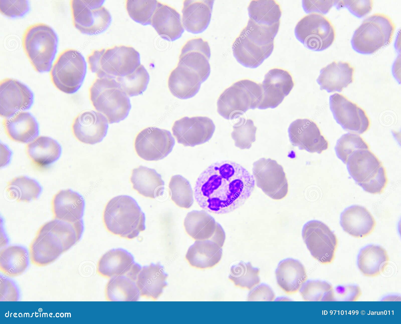
Neutrophil cell stock image. Image of microscope, lymphocyte - 97101499

Live Blood Cell in Darkfield and Phase-contrast Microscopy Course | Cursos  de Medicina Natural
Live Blood Cell in Darkfield and Phase-contrast Microscopy Course | Cursos de Medicina Natural

Neutrophil Show On Blood Smear For CBC Fine Of Microscope. Stock Photo,  Picture And Royalty Free Image. Image 42216403.
Neutrophil Show On Blood Smear For CBC Fine Of Microscope. Stock Photo, Picture And Royalty Free Image. Image 42216403.

Neutrophil cell (white blood cell) in blood smear, analyze by posters for  the wall • posters medicals, leucocyte, erythrocytes | myloview.com
Neutrophil cell (white blood cell) in blood smear, analyze by posters for the wall • posters medicals, leucocyte, erythrocytes | myloview.com

Neutrophil Cell (white Blood Cell) In Peripheral Blood Smear Stock Photo,  Picture And Royalty Free Image. Image 56968571.
Neutrophil Cell (white Blood Cell) In Peripheral Blood Smear Stock Photo, Picture And Royalty Free Image. Image 56968571.

Untitled Document
Untitled Document

LA SANGRE - Pictoeduca

File:Neutrofilo.JPG - Wikimedia Commons

Neutrófilos segmentados, Wright-Giemsa, 1000x. | Download Scientific Diagram

Vacular Stock Photos, Royalty Free Vacular Images | Depositphotos

✓ Neutrophil cell Stock Photos

Leucocitos glóbulos blancos en vena protección contra virus y bacterias el  concepto de ciencia y medicina vista de neutrófilos bajo el microscopio 3d  render | Foto Premium
Leucocitos glóbulos blancos en vena protección contra virus y bacterias el concepto de ciencia y medicina vista de neutrófilos bajo el microscopio 3d render | Foto Premium
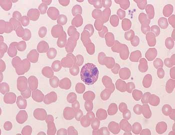
Neutrófilos Segmentados | Llukz | Flickr

Prácticas Inmunología e Inmunopatología (Medicina). Microscopía de las  células del sistema inmunitario
Prácticas Inmunología e Inmunopatología (Medicina). Microscopía de las células del sistema inmunitario

Dos #neutrofilos y un #monocito. #lab #laboratoriodediagnosticoclinico  #hematology #hematología #ciencias #science #leucocitos #microscopio by  lulita_keka http:… | Inspo
Dos #neutrofilos y un #monocito. #lab #laboratoriodediagnosticoclinico #hematology #hematología #ciencias #science #leucocitos #microscopio by lulita_keka http:… | Inspo

Neutrófilos Muestran En La Prueba CBC Frotis De Sangre Se Encontró Con El  Microscopio. Fotos, Retratos, Imágenes Y Fotografía De Archivo Libres De  Derecho. Image 41583423.
Neutrófilos Muestran En La Prueba CBC Frotis De Sangre Se Encontró Con El Microscopio. Fotos, Retratos, Imágenes Y Fotografía De Archivo Libres De Derecho. Image 41583423.

Neutrophil cell (white blood cell) in blood smear, analyze by posters for  the wall • posters medicals, leucocyte, erythrocytes | myloview.com
Neutrophil cell (white blood cell) in blood smear, analyze by posters for the wall • posters medicals, leucocyte, erythrocytes | myloview.com
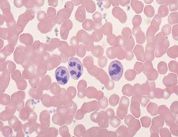
Prácticas Inmunología e Inmunopatología (Medicina). Microscopía de las  células del sistema inmunitario
Prácticas Inmunología e Inmunopatología (Medicina). Microscopía de las células del sistema inmunitario
Jeff (@redb56) / Twitter

Lecture 3 - RBC LAB Flashcards | Quizlet

Un frotis de sangre bajo el microscopio presentan los neutrófilos y  linfocitos rojos. foto micro secciones con alta magnificación con  microscopio de luz Fotografía de stock - Alamy
Un frotis de sangre bajo el microscopio presentan los neutrófilos y linfocitos rojos. foto micro secciones con alta magnificación con microscopio de luz Fotografía de stock - Alamy

Neutrophil Granulocytes Have An Average Diameter Of 12-15 Micrometers In  Peripheral Blood Smears. When Analyzing Neutrophils In Suspension,  Neutrophils Have An Average Diameter Of 8.85 µm Stock Photo, Picture And  Royalty Free
Neutrophil Granulocytes Have An Average Diameter Of 12-15 Micrometers In Peripheral Blood Smears. When Analyzing Neutrophils In Suspension, Neutrophils Have An Average Diameter Of 8.85 µm Stock Photo, Picture And Royalty Free
Blood smear under microscope present neutrophils and red blood cells. Photo  micro sections with high magnification with light microscope Stock Photo -  Alamy
Blood smear under microscope present neutrophils and red blood cells. Photo micro sections with high magnification with light microscope Stock Photo - Alamy